Key Materials for Enhancing 3D Staining Quality
The antibody product line launched by JelloX Biotech is specifically designed for 3D staining, aiming to enhance the precision and stability of pathological research and clinical diagnostics. Our antibodies are rigorously selected for high specificity and sensitivity, enabling precise targeting of proteins within complex thick tissue samples. This ensures clear and consistent staining results while effectively reducing experimental errors.With premium antibodies, users can achieve authentic and reproducible staining outcomes in multi-layered thick tissue views, thereby improving the reliability of digital pathology and immunohistochemistry analysis. JelloX antibodies are suitable for a wide range of tissue samples and research needs, making them a vital tool in advancing pathology and precision medicine.
Detailed product information link: https://jellox.com/antibody/
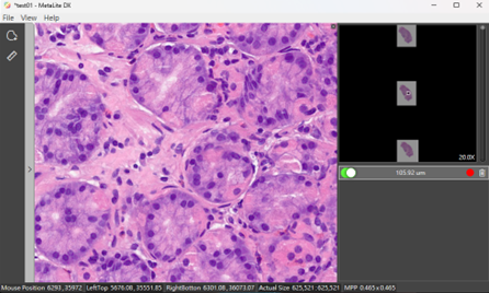
MetaLite DX Digital Pathology Software
MetaLite DX Digital Pathology Software is software designed for viewing digital pathology images. The software is easy to use and intuitive, with many features to help pathologists interpret, diagnose, and manage digital whole slide images. Key features include support for adjusting position, measuring lengths, and adding annotations to specific areas. Note: JelloX Biotech Inc. conducts product security and cybersecurity vulnerability assessment reviews every August until March 07, 2034.
1. Model Number: MLDXUS, Version: 1.2.1, This product has received FDA 510(k) premarket notification approval and been authorized for in vitro diagnostic (IVD) use in the United States.
2.Model Number: MLDXTW, Version: 2.0.0, This product is only sold in Taiwan
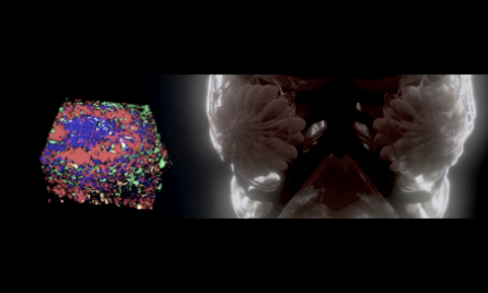
3D Digital Pathology Imaging Service
We provide standard and customized pathology service including tissue staining and clearance, image scanning and processing, and assay report. For example, 3D images of solid tumors such as breast cancer, lung cancer, head and neck cancer with multilayer morphological features or biomarker distribution profiles of clinical grade antibody (Her2, EGFR, PD-L1, Ki67, and CD series, etc.) staining can be achieved in support of precision diagnosis.
2D whole slide image scanning service
2D pathology slide digitization service provides high-throughput, high-resolution and customized image scanning in support of whole slide imaging and important data storage
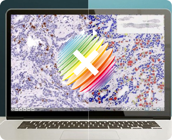
Image data analysis
Nuclear counting
Applying computer vision algorithm to quantify cell staining numbers in pathology images, and make statistical analysis based on cell unit.
Antibody counting
Applying computer vision algorithm to quantify antibody staining numbers in pathology images, and make statistical analysis based on cell unit.
Area analysis
Statistical quantitative analysis of regions of interest based on signal strength of different staining target within annotated pathology image.

AI model
In collaboration with hospitals unique tumor recognition AI models are available for differentiation of normal and cancer cells and facilitation of rapid tumor tissue recognition and analysis
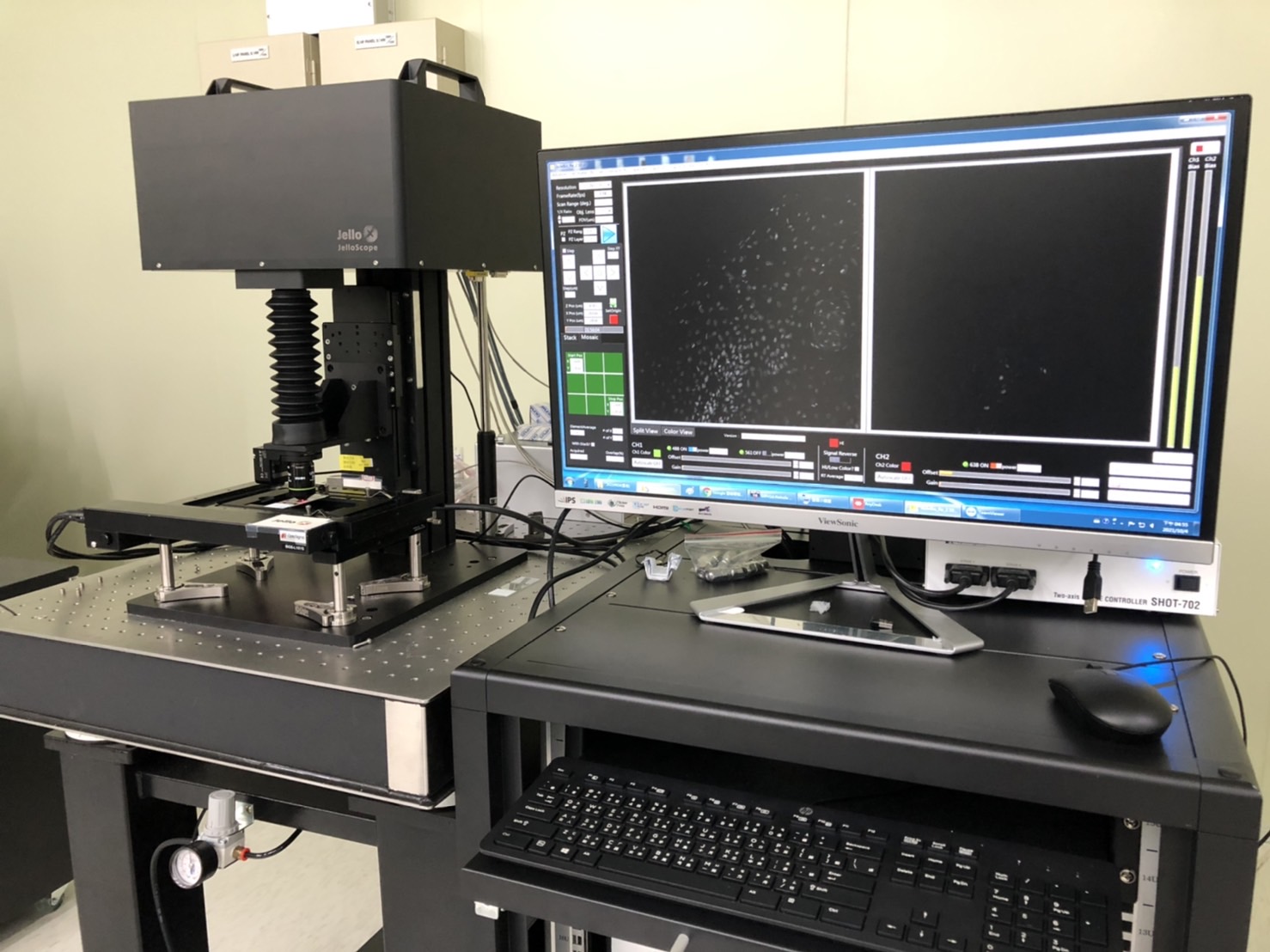
Intelligent pathology image scanner
High speed image scanner JelloScope is well suited for detection of thick pathology biopsies with special advantages of continuously rapid scanning, shortening image acquisition time, and speeding up image processing.

Cloud computation platform
High efficiency cloud computation platform supports value-added service for customers to upload pathology images, select AI software for screening and recognition analysis, and support advanced data analysis.

membrane
staining
antibody
staining
⬤ In clinical setting specific tumor biomarker may be used for cancer screening
antibody
staining
⬤ In clinical setting specific tumor biomarker may be used for cancer screening

products


products